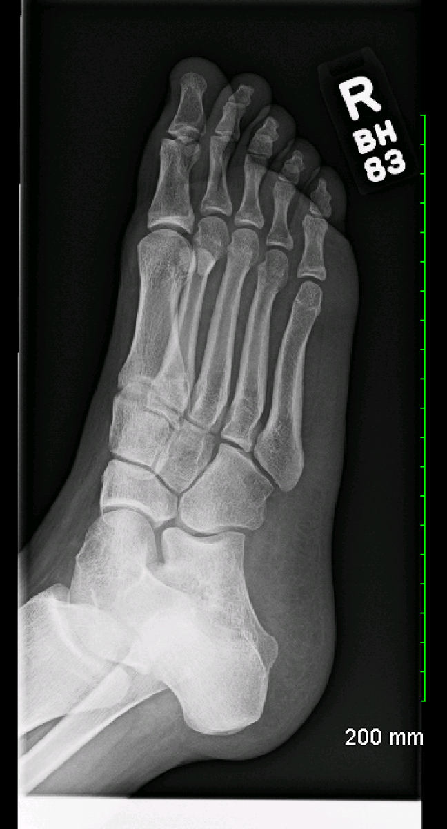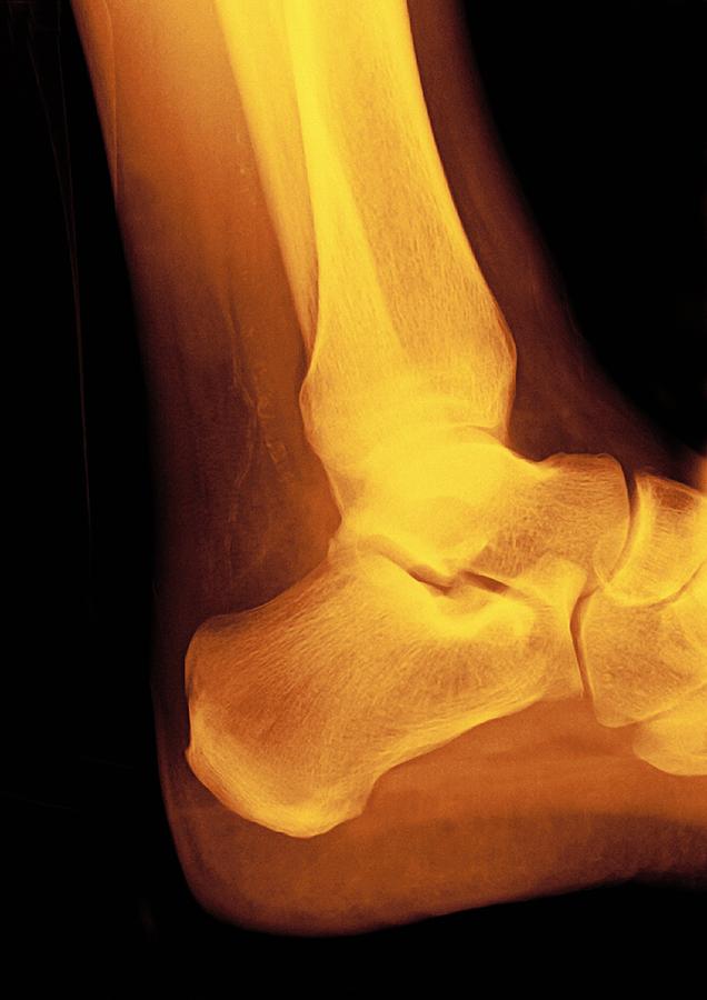

It is always recommended to use standard reference textbooks or published literature. Please understand that this site is not intended to dispense medical advice, provide or assist medical diagnosis. The images on have been quality assured and verified by a senior consultant and specialist in pediatric radiology. Hence the loading times can be slightly above normal, but with zero loss of quality in these normal bone xrays of the children skeleton. The images chosen are unedited and most importantly they are in RAW-format (not compressed). The image is captured on a special X-ray film and is typically in. Injuries of the feet often result in a request for radiographs of the ankle and foot. Normal children chest xrays are also included. X-rays of the foot are completely safe and can show the bones and soft tissues in detail. Procedure Approximate effective radiation dose Comparable to natural background radiation for: Computed Tomography (CT)Brain: 1.6 mSv: 7 months: Computed Tomography (CT)Brain, repeated with and without contrast material: 3. As I and new colleagues constantly had to look up different ossification centers and compare with the present children bone xray at the time – I found having a little library of bone xrays available was very helpful. Extremity (hand, foot, etc.) X-ray: Less than 0.001 mSv: Less than 3 hours: CENTRAL NERVOUS SYSTEM. This site has been made in order to have a quick “reference” look at normal pediatric bone xrays from the ages of day 1 up to 15 years. These normal bone xrays are NOT intended as bone-age references!Įach bone, , represents an image different from the next one, but still within the same localization and age depending on the column and row they are in. Ages are approximate (generally, at most +/- 1-2 months, but mostly within + / – 15 days – unless stated otherwise). Male and female subjects are intermixed. PA films are the standard: the patient stands or sits upright approximately 6 feet in front of the beam source and faces the receptor on the other side, with the X-ray taken while the patient is maximally inspiring (i.e. doi:10.This is a repository of radiograph examples (X-rays) of the pediatric (children) skeleton by age, from birth to 15 years. Journal of the American Podiatric Medical Association. Foot problems in older adults: associations with incident falls, frailty syndrome, and sensor-derived gait, balance, and physical activity measures. Muchna A, Najafi B, Wendel CS, Schwenk M, Armstrong DG, Mohler J. Foot disorders in the elderly: A mini-review. Rodríguez-Sanz D, Tovaruela-Carrión N, López-López D, et al. Potential for foot dysfunction and plantar fasciitis according to the shape of the foot arch in young adults. Flat Foot in a Random Population and its Impact on Quality of Life and Functionality. Pita-Fernandez S, Gonzalez-Martin C, Alonso-Tajes F, et al. Plantar fasciitis in athletes: diagnostic and treatment strategies. Winter foot care: Tips to keep feet warm and cozy all winter long. PMID: 25332943Īmerican Podiatric Association. If the foot is broken it will be put into a cast. All products are produced on-demand and shipped worldwide within 2 - 3 business days. The photograph may be purchased as wall art, home decor, apparel, phone cases, greeting cards, and more. Normalised navicular height truncated H/L). Normal foot x-ray Overview Along with questions of your medical history, your doctor may need to take x-rays of your foot to help aid in making a diagnosis to determine the cause of your foot pain. Normal Foot, X-ray is a photograph by Du Cane Medical Imaging Ltd which was uploaded on May 1st, 2013. The Achilles tendon: fundamental properties and mechanisms governing healing. Normalised navicular height truncated is calculated by dividing the height of the navicular tuberosity from the ground (H) by the truncated foot length (L) (i.e.


X-ray normal humans foot lateral Stock Photo. doi:10.1589/jpts.30.978įreedman BR, Gordon JA, Soslowsky LJ. Find the perfect foot xray stock photo, image, vector, illustration or 360 image. Forefoot transverse arch height asymmetry is associated with foot injuries in athletes participating in college track events.


 0 kommentar(er)
